


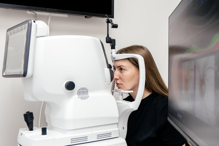
When sponsors ask 20/20 Onsite who should run optical coherence tomography (OCT) across their sites, there are three realistic answers. Certified site staff can scan on site-owned devices, a site's ophthalmology or imaging department can take it on, or a point-of-need ophthalmic partner can bring certified technicians and equipment. Any of the three can work. What really protects the data is whether each scan is taken on a matching device, by a certified technician, following the reading center’s protocol. Below, we compare the three models and what keeps scans consistent in each.
Key Takeaways
- Three models can run OCT across sites: certified site staff, a site's imaging department, or a point-of-need ophthalmic partner, and many trials mix them.
- OCT measurements are not always comparable across devices, so keep each participant on one machine and lock software versions.
- Technicians should be certified to the study's imaging protocol by the reading center, not only trained on the device.
- Reviewing image quality before the participant leaves turns a reading center query into a same-visit rescan.
On This Page
- 1Who Can Perform OCT Scans for Clinical Trial Participants Across Multiple Sites?
- 2How the Three OCT Delivery Models Compare
- 3What Keeps OCT Scans Consistent Across Sites
- 4Where Fundus Photography Fits
- 5Questions to Ask Before Choosing an OCT Provider
- 6How 20/20 Onsite Runs OCT Across Sites
- 7Choose the OCT Model That Fits Each Site
- 8Frequently Asked Questions
Who Can Perform OCT Scans for Clinical Trial Participants Across Multiple Sites?
OCT scans in a multi-site trial are performed by certified technicians at the site, by the site's ophthalmology or imaging department, or by a specialized ophthalmic partner. The reading center grades the images but does not capture them. Sponsors often combine models, matching each site to the option its staff, equipment, and patient volume can support.
The choice matters because OCT drives more than safety checks. A 2024 study in Ophthalmology Retina of 87 retina trials found that 44.5% of screen failures came from patients not meeting imaging criteria. When imaging decides who enrolls, inconsistent scans affect eligibility as well as endpoints.
How the Three OCT Delivery Models Compare
Each model trades setup effort, control, and reach differently. None is right for every site, so the comparison below works best applied site by site.
Comparing OCT Delivery Models Across Sites
| Factor | Trained Site Staff | Site Imaging Department | Point-of-Need Ophthalmic Partner |
|---|---|---|---|
| How It Works | Site coordinators or technicians are trained and certified to scan on a site-owned OCT | The site's ophthalmology or retina department captures scans for the study | A specialized partner brings certified technicians and OCT equipment to the site or to participants |
| Strengths | Scans happen on-site, inside existing visit flow | Experienced imaging staff and established equipment | Matched devices and one certification standard across many sites |
| Watch Points | Training time, staff turnover, and device differences between sites | Department scheduling, and study visits competing with clinical patients | Scheduling coordination with each site's visit calendar |
| Best Fit | Sites with a compatible OCT and stable staff | Academic or hospital sites with retina services | Sites without ophthalmic capacity, or trials needing one standard across sites |
Many trials end up with a mix. A site with a certified retina department keeps its scans in-house, while sites without one use a partner. Our guide to mobile and in-clinic assessment models covers how to match the model to each site.
What Keeps OCT Scans Consistent Across Sites
Consistency comes from three controls, and they apply whichever model a site uses: matched devices and software, certified technicians, and tight alignment with the reading center.
- Matched devices and software: OCT measurements are not always comparable across device models. A 2011 PLOS ONE study found that retinal nerve fiber layer thickness ran 7.4 µm lower on Cirrus than on Stratus. The authors advised against using the two devices interchangeably. They recommended scanning each patient on the same machine throughout a trial. Software version changes can also shift segmentation outputs, so lock versions before the first patient is in.
- Certified technicians: Each technician should be certified to the study's imaging protocol, not only trained on the device. Certification covers scan pattern, centration, signal strength, and how to handle a poor capture.
- Reading center alignment: Settle the imaging charter, file formats, and submission platform before activation. Then review image quality before the participant leaves, so a rescan happens during the visit rather than after a reading center query.
Our guide on BCVA, OCT, and IOP testing in clinical trials covers the operational mistakes that cause OCT drift in more detail.
Where Fundus Photography Fits
Fundus photography follows the same rules as OCT. It captures color images of the retina, and many retina and safety protocols pair it with OCT at the same visit. The same three controls apply: matched cameras, certified photographers, and the reading center's grading criteria. When the protocol calls for ultra-widefield fundus imaging, confirm every site can capture it on a comparable device before activation.
Questions to Ask Before Choosing an OCT Provider
Use these questions with any site, imaging department, or vendor before assigning OCT for a study.
- Which OCT device and software version will be used, and does it match the other sites?
- Are technicians certified by this study's reading center, and how is staff turnover covered?
- Will each participant stay on the same device from baseline through the final visit?
- Is image quality reviewed before the participant leaves?
- How do scans reach the reading center, and who answers its queries?
- Can the provider scale if new sites or regions are added mid-study?
How 20/20 Onsite Runs OCT Across Sites
20/20 Onsite performs OCT and fundus photography through Mobile Vision Clinics, Mobile Vision Pods, and Mobile Clinic Suites, alongside each site's existing capacity. Each model uses calibrated equipment and technicians certified to the study protocol. Clinical teams review images before patients leave the Mobile Vision Clinic, so a poor scan gets retaken during the visit.
The model holds up under pressure. When FDA added ophthalmic assessments midway through four trials, 20/20 Onsite brought 65 sites to full operation in 8 weeks. Each clinic was outfitted with OCT, ultra-widefield fundus imaging, and ETDRS visual acuity testing. A standing relationship with the reading center shortened certification times.
"Sponsors usually ask us who can run OCT. The better question is who can make scan 400 at site 30 look like scan 1 at site 1, and that comes down to devices, certification, and the reading center."
- Dr. Katherine Harkins, Director of Optometry, 20/20 Onsite
Talk to an Ophthalmic Trial Specialist to plan OCT coverage across your sites.
Choose the OCT Model That Fits Each Site
The right OCT plan starts with each site's staff, equipment, and patient volume, then holds every scan to one standard. Whether scans happen in a site's own department, with trained site staff, or through a point-of-need partner, matched devices and certified technicians protect the data. 20/20 Onsite can fill the gaps where sites lack capacity and keep scans consistent across the study.
Schedule a Consultation to map OCT delivery to your sites.
Keep OCT Consistent at Every Site
Talk with 20/20 Onsite about matching each site to the right OCT model, with certified technicians and reading center alignment built in.
Schedule a Consultation